
#Shimadzu multispec 1501 resolution full#
When the cleaning step was completed, the high-pressure vessel (V1) was slowly depressurized and product samples were collected for full physicochemical characterization. The spraying was maintained for 27 min, followed by another 20 min of CO 2 spraying supplied at a rate of 3.0 mL/min to remove any residual organic solvent. When steady-state conditions were reached in the high-pressure vessel (V1 see Fig. 1), the AZT–PLLA solution was released into V1 chamber by a high-pressure pump (P2 see Fig. 1) at a flow rate of 0.75 mL/min and further sprayed through a stainless steel capillary tube (101.6 μm internal diameter). During the production trials, the temperature was maintained at 45 ☌ and the pressure of the system was varied at 85,100, and 135 bar. The schematic representation of the SAS process is depicted in Figure 1.

A bath with ultrasound (frequency 40 kHz, 30 ☌/30 min) was used to remove any gases and homogenize the AZT-PLLA solutions. The AZT–PLLA solution was prepared at 15% (w/v) by dissolving of AZT in 5 mL of ethanol and PLLA in 95 mL of dichloromethane, and then the solution of AZT was mixed with the solution of PLLA. EXPERIMENTAL PROCEDURES Experimental Design X-ray powder diffraction analyses were carried out in X-ray diffractometer from Philips Analytical (X’Pert-MPD Almelo, The Netherlands).

All differential scanning calorimetry (DSC) analyses were carried out in a Differential Scanning Calorimeter from Shi- madzu (DSC-60), equipped with an accessory flow-controller (60A) and an interface for data acquisition and control (TA- 60WS) also from Shimadzu. All infrared spectrophotometric analyses were performed in a Fourier transform infrared (FTIR) spectrophotometer from Thermo Scientific (Nicolet 6700 Madison, Wisconsin) with Fourier transform, coupled with an Omni Smart-Sampler also from Thermo Scientific. Scanning electron microscopy analyses were carried out in a scanning electron microscope (SEM) from Leica (Leo 440i Cambridge, England), following gold sputter coating of the particles with a Sputter-Coating plating device also from Leica (SC7620). All dissolution tests were carried out using a dissolution apparatus from American Lab (AL1000 Charqueada, Brazil).

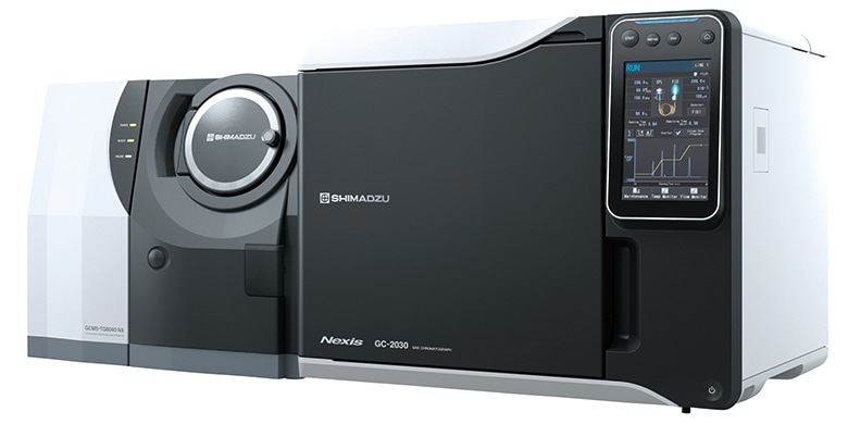
All spectrophotometric readings were carried out in a UV–Vis spectrophotometer from Shimadzu (MultiSpec-1501 Kyoto, Japan). For dissolution and the removal of the dispersed gas in solution was used bath ultrasound (Unique USC2800A Indaiatuba, Brazil). The SCF system utilized for this research work was from Waters Corporation (SAS-RESS Combined System, Pittsburgh, Pennsylvania). It has been demonstrated that it is possible to use PLLA carrier and SAS process to obtain SD, in a single step. AZT release is controlled by a diffusion mechanism. AZT remained in crystalline form, whereas PLLA remained in semicrystalline form. Intestinal permeability studies using the AZT–PLLA from 元 batch led to an AZT permeability of approximately 9.87%, which was higher than that of pure AZT (~ 3.84%). The 元 batch of SD, produced with 1:2 (AZT–PLLA) ratio, resulted in a 91.54% yield, with 40% AZT content. From the nine possible combinations of tests performed experimentally, only one combination did not produced a solid. AZT–PLLA production batches were carried out by the SAS process, and the resulting products evaluated via scanning electron microscope, X-ray diffraction, differential scanning calorimetry, and Fourier transform infrared analyses. A 3 2 factorial design was used, having as independent variables the ratio 3′-azido-2′3′- dideoxythymidine (AZT)–PLLA and temperature/pressure conditions, as dependent variables the process yield and particle macroscopic morphology. A supercritical antisolvent (SAS) process for obtaining zidovudine-poly( l-lactic acid) (PLLA) solid dispersions (SDs) was used to attain a better intestinal permeation of this drug.


 0 kommentar(er)
